


Orthopedic surgeons focus on the bones, joints, muscles, ligaments, and tendons that keep you moving. In sports medicine, they identify what went wrong, how it happened, and what structures are involved. Their approach is systematic and grounded in evidence. It moves from your story to hands‑on testing to targeted imaging. By following a clear process, they narrow the diagnosis and guide next steps with accuracy and care.
Reviewing Symptoms & Medical History
First comes your account of the injury, and it sets the direction for everything that follows. You describe the sport, the exact moment symptoms began, and whether there was a pop, twist, or direct blow. Orthopedic surgeons may ask about the location of pain, the timeline of swelling, locking, instability, numbness, and weakness. They also review training load, footwear, playing surface, and past injuries that might influence current risk, which helps link patterns to likely structures.
A detailed timeline helps to separate acute tears from overuse problems. Medication use, prior surgeries, and systemic conditions, such as diabetes or autoimmune diseases, can affect tissue healing and require caution. While brief, targeted questions about work duties and school sports calendars help map functional demands; this context shapes the diagnostic path.
Providing Physical Exams
They observe first, then they touch, and finally they stress specific tissues. The exam begins with an assessment of gait and posture. Swelling, bruising, deformity, and muscle atrophy are noted. Tests are used to pinpoint tenderness along ligaments, bony landmarks, or tendon insertions. Range-of-motion testing may compare sides to detect stiffness or laxity.
Strength testing with resistance spots deficits from pain inhibition versus true muscle or nerve injury. Other checks review sensation, reflexes, and pulses. Findings are interpreted together, not in isolation, because single tests may lack perfect accuracy.
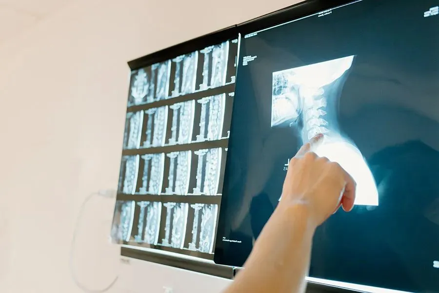
Using Imaging Techniques
Imaging refines the picture, and it does so with tools chosen for the question at hand. X‑rays show fractures, joint alignment, and growth-plate status in adolescents, while they may look normal in pure soft‑tissue injuries. Ultrasound offers dynamic views of tendons and ligaments, detects fluid collections, and guides procedures; it is quick, portable, and effective for superficial structures. MRI provides high‑contrast detail and helps stage injury severity and map exact locations. CT supplies 3D bone detail for complex fractures. Bone scans and advanced MRI sequences may help in stress injuries or unusual fractures.
Imaging is interpreted alongside history and exam rather than as a standalone answer. An MRI that shows signal in a tendon aligns with palpation tenderness and load pain during resisted testing. When results conflict with clinical findings, surgeons repeat targeted maneuvers, adjust hypotheses, or order alternate views to resolve uncertainty.
Get Help From Orthopedic Surgeons
Diagnosis guides safe activity choices, training adjustments, and timing for recovery steps, so getting it right matters. If you have persistent pain, swelling, instability, or loss of function after a sports incident, schedule an evaluation with an orthopedic surgeon. Bring details about the onset, training changes, and any prior injuries; wear clothing that allows joint access; and note what movements trigger symptoms. Take the next step today and reach out to an orthopedic specialist to get a precise diagnosis and a plan according to your activity demands.


Leave a Reply