


Ultrasound and MRI provide different views of the body during urgent evaluations. Ultrasound uses sound waves to produce real-time images, which support quick assessments of fluid, blood flow, and organ movement. MRI uses magnetic fields and radio waves to reveal detailed soft-tissue structures, nerves, and subtle injuries. Here’s how teams choose ultrasound vs MRI applications in emergency care:
1. Trauma
First, teams run a focused assessment with sonography for trauma (FAST). This bedside ultrasound survey checks for free fluid in the abdomen, pelvis, and around the lungs or heart. While brief, the exam provides immediate feedback, and repeating it during resuscitation allows for tracking changes. Extended FAST adds lung views to help identify potential issues.
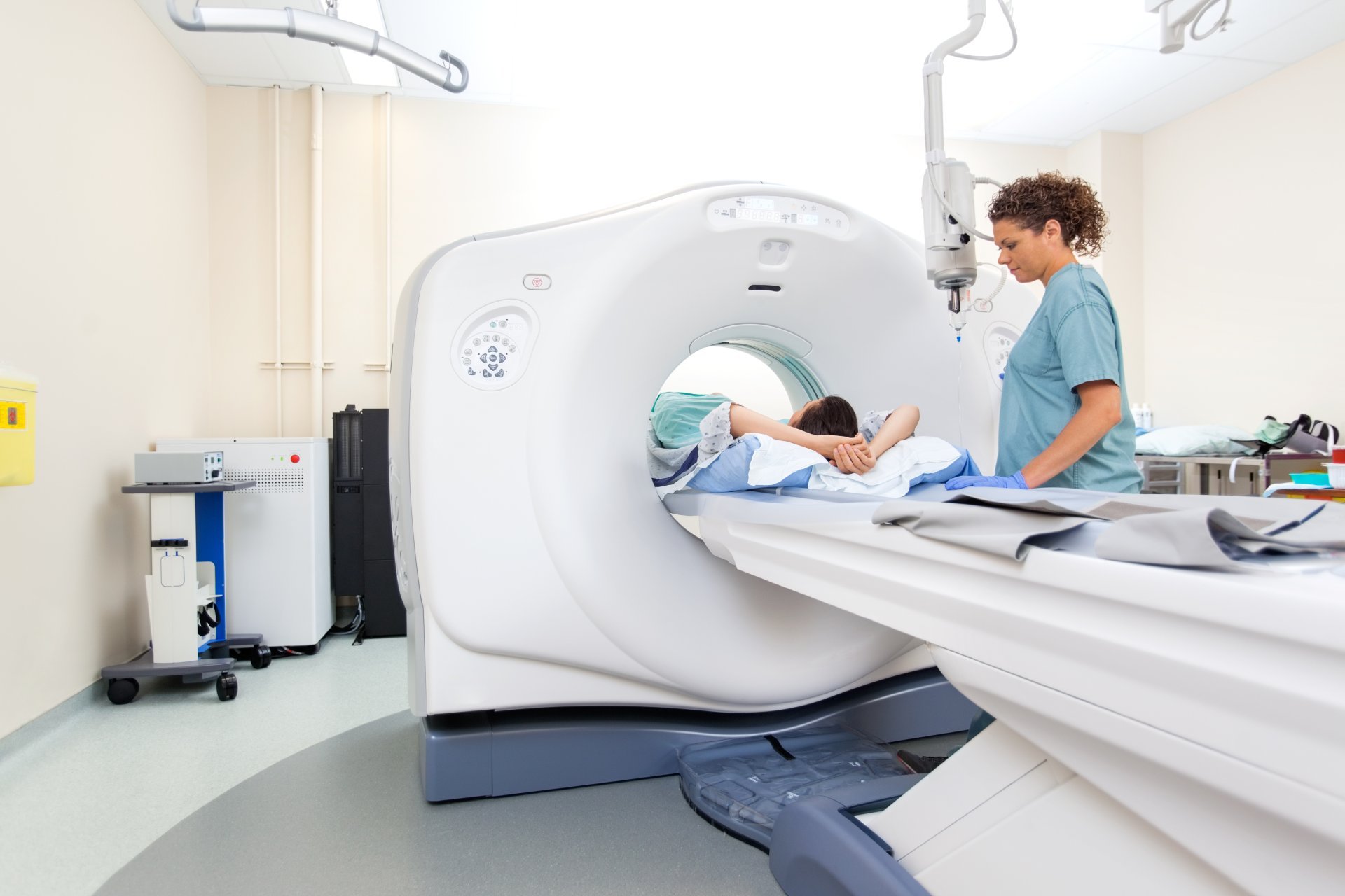
MRIs are used when detailed information about soft tissue is required. It maps ligament tears after joint injuries, evaluates spinal cord issues, and characterizes certain fractures that are not visible on initial X-rays. While not used during active resuscitation, MRI helps refine the diagnosis once the patient is stable, and it guides plans with high-contrast views of muscle, tendon, and marrow.
2. Cardiac Arrest
During cardiac arrest, teams carefully plan procedures, and an ultrasound may be used during short pauses in chest compressions. A quick scan from below the chest or beside the heart can show if the heart is not beating properly or if there is abnormal fluid around it, and can also check if the right side of the heart is enlarged, which might suggest a blood clot in the lungs. These simple scans help guide checks for a pulse and assign tasks to team members, without stopping chest compressions; a planned time for imaging keeps the care flow steady.
After professionals restore the person’s heartbeat, and their neurological condition is still uncertain, MRI scans can be helpful. A brain MRI can reveal areas damaged due to a lack of oxygen. Special MRI techniques can look for blockages or tears in major blood vessels if a stroke is suspected. More detailed MRI scans can also help detect heart muscle inflammation or scarring, which can aid in determining the right long-term treatment.
3. Abdominal Pain
Doctors use an MRI when radiation is avoided or soft-tissue contrast is needed. Pregnant patients with right lower pain may receive an MRI to evaluate appendicitis without ionizing radiation. Doctors may prefer this imaging option when a clearer view of soft tissues is required, as it provides detailed and accurate results. An MRI can also be a safer choice for individuals who should avoid radiation exposure. This may include children and pregnant individuals when choosing between ultrasound vs MRI methods.
4. Pelvic Issues
Pelvic issues vary depending on a person’s age and gender, and imaging tests should match the body’s anatomy, symptoms, and how urgent the situation is. Professionals use ultrasounds early in pregnancy to check for bleeding; they can find various pregnancy issues. Quick scans can evaluate bladder size, blood buildup in the uterus, or fluid in the pelvis after an injury. MRI scans may provide clearer images for specific medical conditions.
Choose Ultrasound vs MRI Imaging
Match each question to the appropriate tool and plan the next step accordingly. Use ultrasound for quick checks and guided procedures. Turn to MRI when detailed tissue characterization, brain assessment, or comprehensive mapping can influence your decision. Both imaging methods have distinct roles and are most effective when chosen based on the clinical question. For a more detailed guide on protocols, indications, and common findings, visit our resource hub or contact an imaging specialist to discuss your case.


Leave a Reply